Surgical Technique Guide for Wrist Hook Volar
Alexander Y. Shin, M.D.
Professor and Consultant of Orthopaedic Surgery
Department of Orthopaedic Surgery, Division of Hand Surgery
Mayo Clinic, Rochester, MN
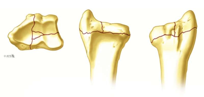
The indications for the Volar Hook Plate include intra-articular volar ulnar and volar radial distal fracture fragments that cannot be held securely by volar plate techniques. These fragments are typically within 3-5 mm from the volar articular margin and distal to the watershed line. Illustrated here is a typical complex intra-articular fracture with a die punch fragment, dorsal ulnar fragment, volar ulnar fragment and radial styloid fragment.
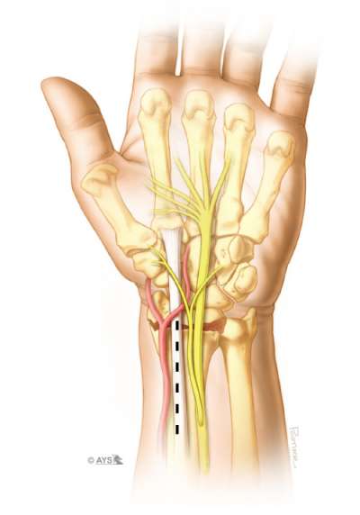
A volar approach to the distal radius is made with an incision that follows the flexor carpi radialis tendon. Rarely is the incision made distal to the wrist crease, however, surgeon preference will dictate length of the incision. If a multi incision approach is made to the wrist, the incision can be shifted ulnarly by 0.5cm to allow enough of a soft tissue bridge between the radial incision (for the radial styloid fixation). Alternatively, the radial styloid can be fixed with an extended FCR incision by angling the distal portion of the incision radialward
The flexor carpi radialis tendon is mobilized, the floor of the flexor carpi radialis is incised and the flexor tendons/median nerve are retracted ulnarward. The brachioradialis tendon is identified and divided if necessary to facilitate fracture reduction.
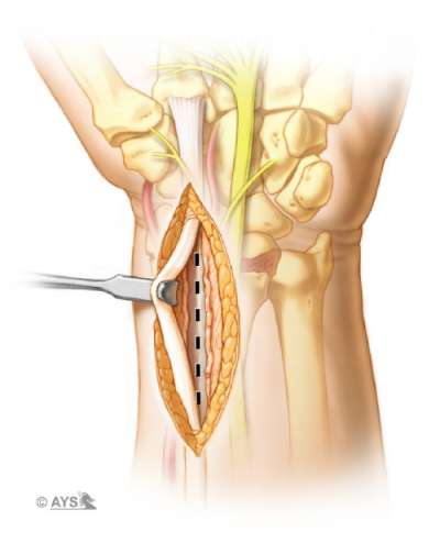
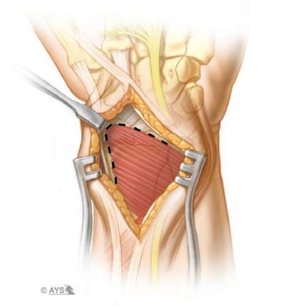
The pronator quadratus muscle is identified and the radial and distal margins are visualized. The pronator quadratus is sharply elevated from the radial side of the radius and bluntly elevated from the distal margin, taking care not to divide the radiocarpal ligaments. It is important to elevate the pronator quadratus to the most ulnar aspect of the radius.
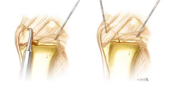
Intra-articular reduction may be further adjusted by using limited periarticular incisions to allow: direct manipulation of articular fragments; placement of subchondral bone graft; repair of intercarpal ligament injuries; and augmentation of fractures with K-wires, Buttress Pins, Hook Plates or Pin Plates. Displaced volar ulnar corner fragments that are not reduced with the Bridge Plate alone require buttress support.
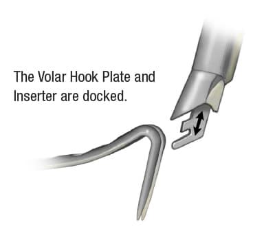
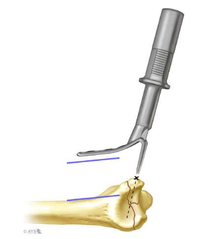
The plate and Inserter are held such that the tines of the Hook Plate are placed at approximately 1-2 mm from the distal edge of the radius. The shaft of the Hook Plate is positioned parallel to the radius shaft. Because the tines of the Hook Plate are angled back, they will engage into the fragment and not be intraarticular. Flouroscopic image of a perfect lateral of the radius can be of assistance.
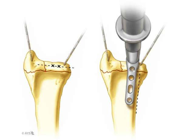
For a Hook Plate placed ulnarly, the tines are positioned parallel to the articular surface, and the radio-ulnar alignment is also verified so the ulnar edge of the plate will be collinear with the ulnar aspect of the radius shaft. The Inserter is gently tapped with a small mallet and the tines are engaged and tapped into the fracture fragment. Just prior to seating the Hook Plate fully, the Inserter is disengaged and reposited over hooks for final impaction.
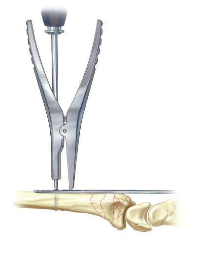
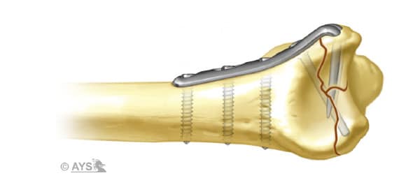
Holding the proximal aspect of the plate to the radius, the 1.8 mm drill is used to secure the plate to the radius shaft with a 2.3mm screw. A locking peg (either threaded or smooth) can then be placed in the locking hole when the dorsal fragments have been reduced. Verification of placement on fluoroscopic image is necessary. An additional Hook Plate can be placed radially if needed.
Disclosure: The author did not receive any outside funding or grants in support of this work. Neither he nor a member of his immediate family received payments or other benefits or a commitment or agreement to provide such benefits from a commercial entity.